Skip to content
+605-246 1977
+605 – 241 9000
info@pcsh.com.my
Results
Home
About
Vision & Mission
Our History
Our Team
Facilities & Services
24-Hour Accident & Emergency
Operating Theatres
Haemodialysis Centre
Imaging Centre
Pharmacy
Dietetic Resource Centre
Wards
Heart Centre
Diagnostic Centre
Wellness Centre
Physiotherapy Centre
Patient Relations
Other Facilities
Doctors
Our Consultants
Anaesthesiologists
Cardiologist Physician
Dermatologist
Ear, Nose & Throat (ENT) Surgeons
General Surgeons
Nephrologists & Physician
Neurologist & Physician
Obstetrician & Gynaecologists
Ophthalmologists
Orthopaedic Surgeons
Paediatrician
Paediatrician & Paediatric Neurologist
Physicians
Plastic & Reconstructive Surgeon
Psychiatrists
Radiologists
Respiratory & Physician
Rheumatologist & Physician
Urologists
Visiting Consultants
Visiting Consultants
Patient & Visitors
Health Screening
Career
Contact
Imaging Centre
Contact Us
+605 - 246 2964
Contact Now
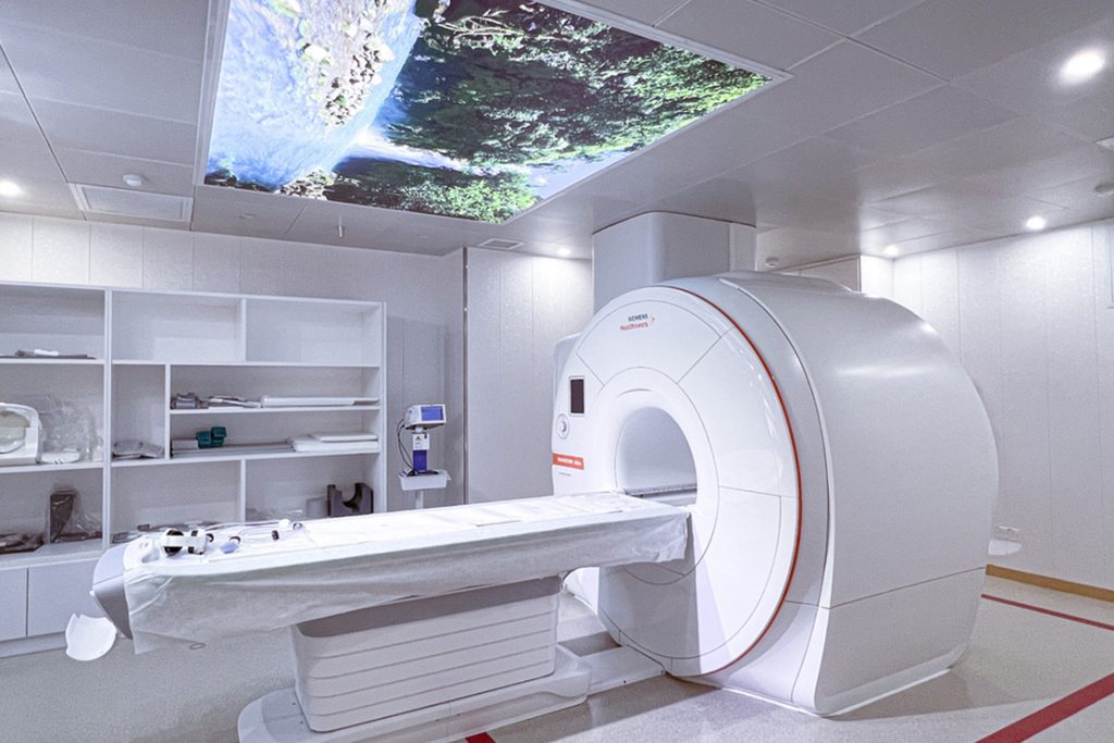
Magnetic Resonance Imaging (MRI)
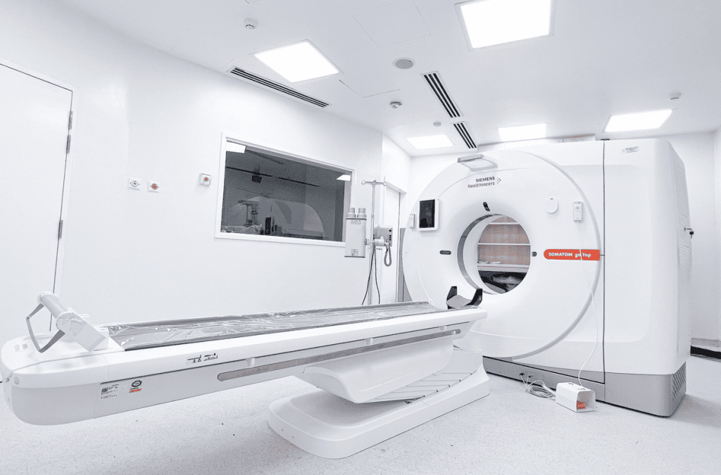
Computed Tomography (CT) Scan
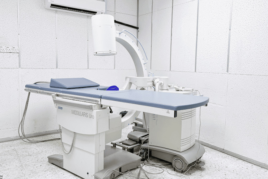
Fluoroscopy
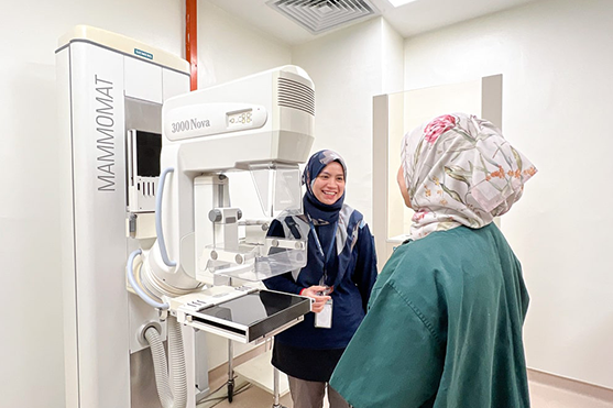
Mammogram
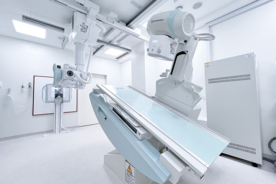
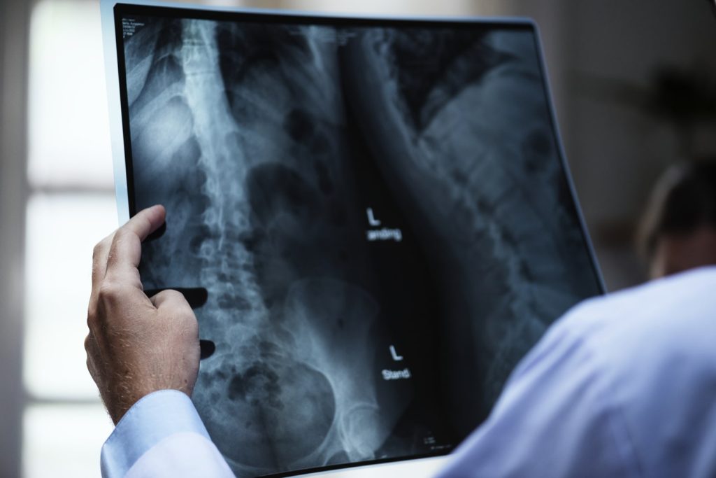
X-Ray
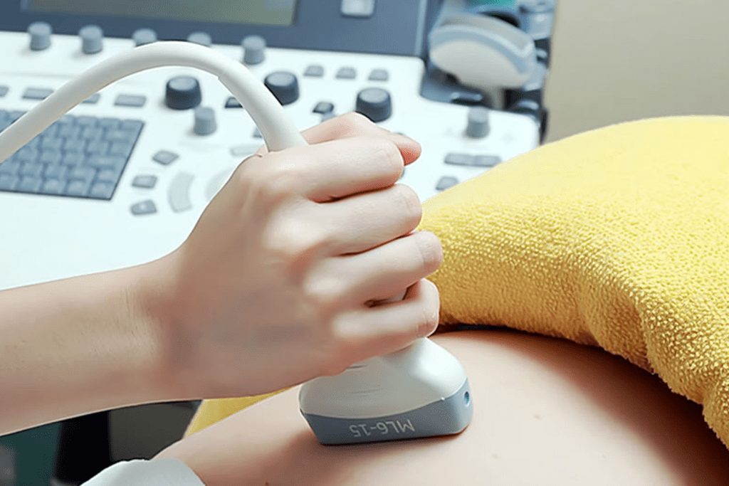
Ultrasound
Lithotripsy Centre